

Dolphin Imaging 3D Surgery
Advanced software module designed to streamline orthognathic surgical planning and treatment through comprehensive 3D imaging and simulation
Input Data Compatibility
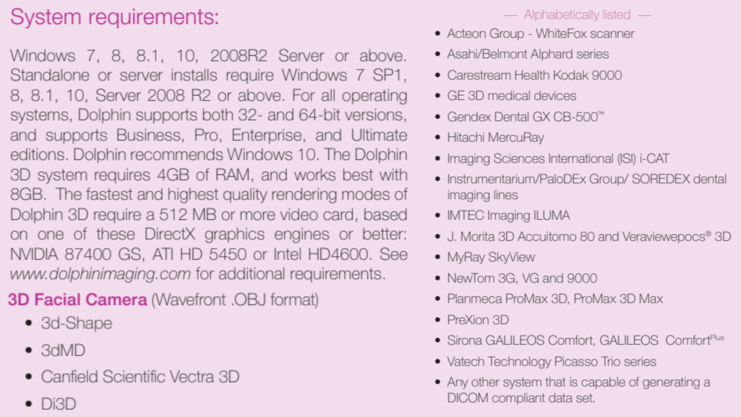
- High-resolution 3D cone beam or spiral CT images serve as the primary data source.
- Optional augmentation with 2D or 3D facial photographs and laser or optical scans of dental models.
- Supports DICOM 3 standard (multi-file or single file) from a wide range of CT and CBCT scanners (see Compatibility Table below).
- Compatible with 3D facial cameras producing Wavefront .OBJ format.

Comprehensive Surgical Planning Tool – “Treat”
- Enables planning from multiple views: lateral, frontal, and submento-vertex (SMV).
- Provides numerous clinical and numeric tools tailored for Surgeon, Technicians and Researchers
- Facilitates quick preliminary workups, interactive case discussions, or highly detailed surgical plans.
- Allows simulation of skeletal movements and real-time soft tissue contouring Profile adjustments, Frontal nasal region shaping and Lip region modifications
- Displays soft tissue changes dynamically during surgical manipulation.
Animated Treatment Presentation – “Present”
- Converts the simulated surgery plan into animated sequences showing pre- and post-operative skeletal and soft tissue configurations in 3D.
- Useful for Detailed study of treatment nuances, Patient education and consent and Case presentations to surgical teams
- Advanced tools allow airway analysis along curved paths, quantifying airway volume and cross-sectional areas with color-coded constriction visualization.
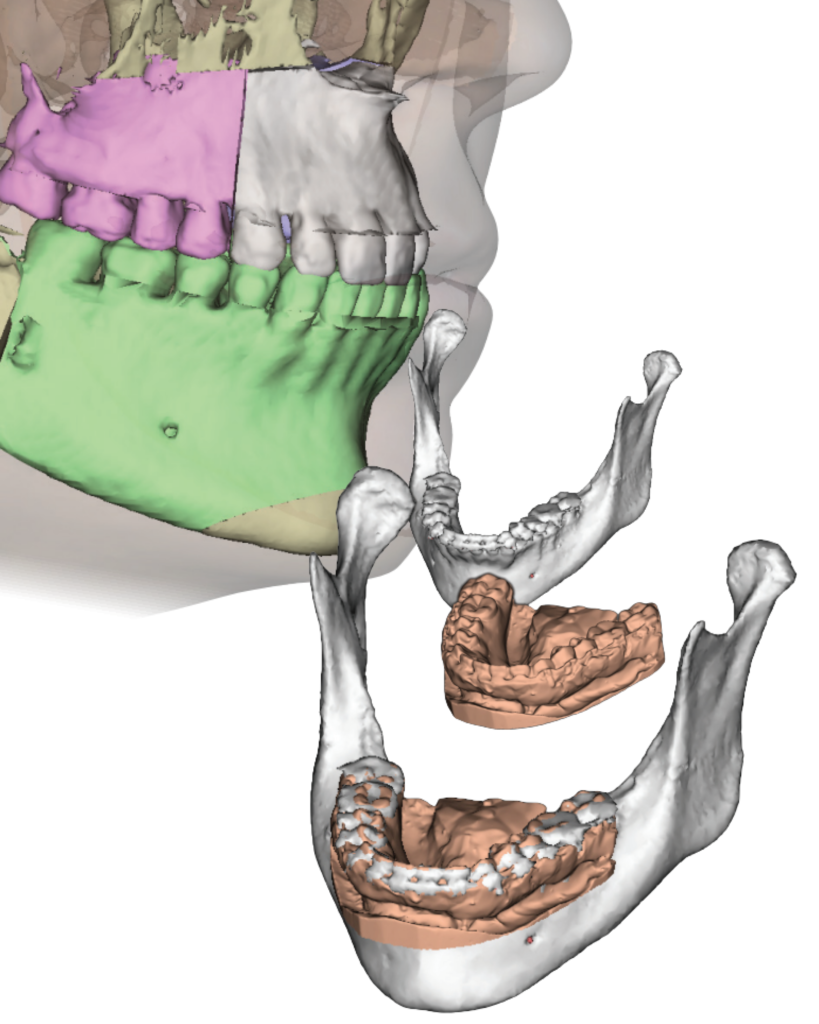
Customizable Surgical Guide Fabrication – “Splint” Tool
- Customizes intermediate and final splints considering width and thickness.
- Supports maxillary and mandibular positioning based on the operational sequence.
- Outputs splint data in industry-standard .STL format.
- Compatible with 3D printers or external lab manufacturing.