

Dolphin Imaging 3D
FDA-cleared Class II medical device designed for specialized dental practices and research institutions. It facilitates comprehensive 3D visualization, diagnosis, treatment planning, and documentation of craniofacial anatomy
Dolphin Imaging 3D Software Overview
- Dolphin Imaging software is an FDA-cleared Class II medical device designed for dental specialists to capture, analyze and present craniofacial images.
- It processes data from CBCT, MRI, medical CT, and 3D facial camera systems, supporting diagnosis and treatment planning across multiple dental disciplines.
- The software depends on interpretation by trained and licensed practitioners, emphasizing its use as a professional diagnostic tool.

User Interface and Accessibility
- Dolphin 3D features a simple, intuitive GUI that enables easy manipulation of complex volumetric datasets without requiring knowledge of scripting or commands.
- The software supports importing a variety of 3D dataset formats and offers versatile visualization options including 3D volume rendering, cross-sectional views, and 2D photo wrapping.
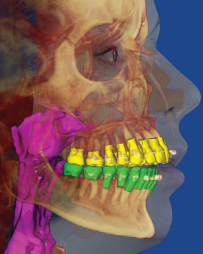
- Users can segment and visualize anatomical structures by adjusting intensity levels for soft and hard tissues, translucency, and color coding.
- Enhanced object orientation tools allow users to establish consistent default orientations based on anatomical planes (mid-sagittal, axial, coronal) and adjust yaw, pitch, and roll.
- Multiple planar layouts facilitate simultaneous viewing of 3D volumes and orthogonal cross-sections.

Measurement and Analysis Capabilities
- The software supports 3D and 2D measurements of distances, angles, and areas with digitized landmarks that can be exported for further analysis.
- It includes cephalometric and panoramic radiograph generation from 3D datasets using customizable projection methods.
- Advanced tools allow airway analysis along curved paths, quantifying airway volume and cross-sectional areas with color-coded constriction visualization.
Advanced Imaging and Reporting Features
- Dual volume superimposition enables comparison of scans from different timepoints to track treatment progress accurately.
- The Hounsfield Unit (HU) color mapping tool differentiates biological tissues by radiolucency for detailed structural analysis.
- The software supports 2D facial photo wrapping to texture-map patient facial photographs onto 3D volumes, improving case presentation realism.
Additional Functionalities and Integration
- Automated movie scripts allow users to create animated visualizations of 3D data with customizable parameters such as position, zoom, and translucency.
- Diagnostic reports can be generated using customizable templates tailored to different dental specialties and clinical indications.
- Digital Study Model software supports integration with intraoral and study model scanners, compatible with STL and OBJ file formats, and works with CEREC systems for orthodontic and restorative workflows.
